| | | | | | |
| The Brain |
Figure 2: a sagittal section through the human brain showing the structures of the left side.
The labels for Fig.2 are as follows:
3, 4
The third and fourth ventricles – cavities filled with circulating cerebrospinal fluid (CSF).
AC = anterior commissure
A white fibre bundle connecting the two cerebral hemispheres.
AV = arbor vitae (‘tree of life’)
White fibre tracts in the cerebellum, carry information between cerebellum and thalamus, brainstem and cerebral cortex.
C = colliculi (part of tectum or roof of the midbrain)
Inferior colliculi – one pair of nuclei for relaying auditory inputs from pons (cochlear nerve → cochlear nucleus →
superior olive in pons) to thalamus;
Superior colliculi – one pair of nuclei for relaying visual inputs from visual cortex and retina to thalamus (controls eye
movements, e.g. saccades).
CA = cerebral aqueduct (aqueduct of Sylvius)
Connects the third ventricle to the fourth ventricle, contains cerebrospinal fluid (CSF).
CC = corpus callosum
The main white fibre tract connecting the two cerebral hemispheres, arc-shaped, contains 10 x more fibres than the
anterior commissure.
CG = cingulate gyrus
The innermost gyrus down in the longitudinal fissure above the corpus callosum (one pair of these, one on each
hemisphere) – part of the cingulate cortex which is part of the limbic system governing emotions, drive and olfactory
memory – the CG is implicated in hypnosis.
CP = choroid plexus
Modified regions of ependymal cells (cells lining the ventricles) which secrete CSF (absent from CA) – secrete about 500
ml CSF / day.
F = fornix
A pair of arc-shaped fibre tracts which connect the limbic system in each hemisphere – connect the hippocampus (x2) to
the hypothalamus then to the mammillary body (x2) and then to the thalamus (x2).
H = hypothalamus
Region beneath the thalamus, controls much of the endocrine system via the pituitary gland; regulates sweating, thirst,
hunger, blood pressure and heart rate; contains the suprachiasmatic nucleus (biological clock controlling circadian
rhythms); essential for the coordination of emotional responses.
MB = mammillary body
One pair of these, part of limbic system (‘instinctive brain’ controlling emotions, ‘animal’ drives and the formation of long-
term memories, especially emotional ones which have a large olfactory component).
MI = massa intermedia
A bridge of grey matter connecting the pair of thalamic masses together (see thalamus).
MO = medulla oblongata
Contains motor and sensory tracts connecting the spinal cord to the pons; posterior surface forms the floor of the 4th
ventricle; regulates certain reflexes: coughing, sneezing, swallowing and vomiting; cardiac centre regulates heart rate,
vasomotor centre regulates blood pressure; vestibular nuclei receive inputs from vestibular nerves and connect to the
cerebellum; reticular activating system (part of reticular formation) in medulla, pons and midbrain form an arousal system
(damage to which can result in coma).
OB = olfactory bulb
One pair of these, enlarged terminus of olfactory nerve, gives out sensory fibres into the nasal cavity through the porous
cribriform plate.
OC = optic chiasma
Region where some of the fibres in the pair of optic nerves cross-over before reaching the thalamus.
PG = pineal gland
Regulate circadian rhythms by secreting melatonin (controlled by the suprachiasmatic nucleus in the hypothalamus.
Pi = pituitary gland
The ‘master gland’ regulating the endocrine system.
Pons (pons Varolii)
Literally ‘bridge’ – connects the medulla oblongata to the cerebellum and the medulla to the midbrain; helps the medulla
regulate respiration (contains a nucleus essential for the switch between inspiration and expiration).
SP = septum pellucidum
A sheet of tissue between the pair of lateral ventricles.
T = thalamus
A paired structure (fused together via massa intermedia); primarily acts as a ‘switchboard’ to relay signals from sensory
tracts in the brainstem to the appropriate parts of the cerebral cortex; form part of the walls of the third ventricle which they
surround; helps regulate consciousness, sleep and alertness; helps regulate pain; triggers brain waves in the cerebral
cortex.
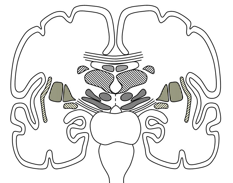
Figure 3: coronal section through the human brain, illustrated the basal nuclei and associated structures.
The Basal Nuclei / Ganglia
The cross-section of the human brain, Fig. 3, which is a coronal section (from 'ear to ear') illustrates the basal nuclei and
associated structures. A ganglion is a coordinated mass of nerve cell nuclei and a centre of information processing. Strictly,
ganglia occur as swellings on nerves outside the CNS and ganglia occurring inside the CNS should be referred to as nuclei.
C: claustrum
A sheet of white and grey neurones of unknowbn function, but implicated in communication between cortical areas involved in
attention.
CN: caudate nucleus
A pair of C-shaped structures (with its head and tail pointing forwards / rostral). Along with the putamen, the caudate nucleus
receives inputs from the thalamus, cerebral cortex and brainstem.
F = fornix
A C-shaped white fibre tract, which connects the hippocampus (Hi) to the hypothalamus.
GP = globus pallidus (pallidum)
The output nucleus of the basal ganglia, sends its outputs to the thalamus.
H = hypothalamus
The hypothalamus has a variety of functions but one of its most critical is the regulation of the endocrine 9hormonal
system) via the pituitary gland.
Hi = hippocampus (pl. hippocampi)
A pair of deep infoldings of the cerebral cortex of the temporal lobe, there is one of these horizontal
seahorse-shaped structures on either side of the brain. Sometimes said to be underneath the cortex, but strictly an
ancient part of the cortex (part of the allocortex, lit. 'other cortex', which evolved before the much larger neocortex, lit.
'new cortex'). The hippocampi are important for spatial memory and for converting short-term memories into
long-term memories, but long-term memories are not generally stored here, so damage to this region of the brain
impairs the ability to form new memories but not the retrieval of old memories. Part of the limbic system, which is
concerned with emotions, the hippocampus is especially important in processing emotional memories and
converting these into long-term memories. Of special importance, in this regard, are olfactory memories. Have you
ever walked into a room and noticed a familiar smell which reminded you of some experience or feeling you
associated with the same small a long time ago? This is the work of the limbic system. The association of smells
with emotions results in the formation of very persistent memories.
LN = lenticular nucleus
The globus pallidus and the putamen.
SN = substantia nigra
Part of the midbrain, consists of an output region (pars reticulata) and a dopamine synthesising / storing region (pars
compacta). The dopamine gives the substantia nigra a dark colour. The substantia nigra are part of the midbrain or
mesencephalon. The midbrain also contains a pair of red nuclei which receive inputs from the cerebellum and relay
outputs to the spinal motor tracts (rubrospinal tracts).
Striatum
Consists of the caudate nucleus, putamen and ventral striatum (nucleus accumbens).
Function of the basal nuclei
Inputs from cerebral cortex, thalamus and brainstem enter the basal nuclei via the caudate nucleus and putamen. Outputs from
the globus pallidus pass to the thalamus and brainstem. The thalamus relays outputs to the motor cortex of the cerebrum.
The prime function of the basal nuclei is to regulate the activity of the motor cortex via thalamus, by means of feedback loops,
to ensure smooth muscle movements. The substantia nigra modifies the outputs from the pallidum by releasing dopamine as a
neurotransmitter. The output from the basal nuclei inhibits the thalamus and hence also the motor cortex. Dopamine regulates
this output by reduces the basal nuclei output strength which increases thalamic and cortical activity. Thus, the balance
between the dopamine output of the substantia nigra and the output of the pallidus determines the correct level of feedback to
the thalamus. In Parkinson's disease, degeneration of the substantia nigra means that the output strength of the basal ganglia
is too strong and the thalamus - motor cortex circuit is excessively inhibited. This means that sufferers of this condition have
difficulty initiating movements (their movements are over-inhibited). For this reason, Parkinson's disease is a hypokinetic
disorder (i.e. a disorder characterised by inhibited movements). This basal nuclei circuit is illustrated below:

Figure 1: surface features of the human brain (left hemisphere). The human brain is a highly sophisticated
computer consisting of neurons and accessory cells called neuroglia and other supporting tissues. Terms like
anterior and posterior are a bit ambiguous when describing the brain of a biped like the human being, so we
have used rostral (R) for the noseward pole, and caudal for the pole pointing to the back of the head.
The human brain is a very powerful biological computer consisting of about 100 billion (10^11 neurons, the
actual number is thought to be closer to 86 billion) and about ten times as many glial cells. The glial cells
provide mechanical and immune protection for the brain as well as performing several other functions such as
regulating irrigation of the brain for waste removal and providing electrical insulation to neurone axons (the
'wires' of the brain). The neurones are involve din information processing, although the glial cells possibly also
have a role in this. The neurones are connected by electrical and chemical synapses, with a pyramidal cell in
the cerebral cortex having about 35 000 connections to other neurones! The whole forms a vastly complex
information-processing network. Most of this information is processed subconsciously (unconsciously). The
conscious mind may have problems calculating simple mathematics, but make no mistake, the brain as a
whole performs a tremendous number of computations every second! This is why you can catch a ball without
thinking too much about it! You don't need to sit there and draw a diagram of the trigonometry and solve the
velocity vectors as the ball whizzes towards you, because luckily this is done automatically by this very
powerful computer!
Computers are energy-consuming, and the brain requires about 15% of the cardiac output in blood flow and
uses about 20% of the body's energy. Thinking uses energy! To date (2014) the most powerful
supercomputers on Earth (using almost one million processor cores) require about 40 minutes to emulate one
second of activity in 1% of the human brain! The human brain is still the most powerful computer on Earth
(which makes it all the more tragic that so many are mis-used!). The human brain carries out an estimated
10^18 computations per second. Electronic digital computers are expected to reach this level in a few years
time, however.
The brain is divided into the forebrain (prosencephalon) the midbrain and the hindbrain. The forebrain
consists of the two cerebral hemispheres, or telencephalon, and the thalamus (paired structures) or
diencephalon.
The midbrain (mesencephalon) is divided into two main regions:
1) the ‘roof’ or tectum of the midbrain, posterior (caudal) to the cerebral aqueduct. The colliculi are found
here. The superior colliculi are also called the optic tectum. The four colliculi (one inferior pair and one
superior pair) are also called the corpora quadrigemina.
2) The ‘floor’ or tegmentum of the midbrain, anterior (rostral) to the cerebral aqueduct. The red nuclei and
substantia nigra are found here.
The hindbrain (rhombencephalon) consistas of the metencephalon (pons and cerebellum) and the
myelencephalon (medulla oblongata).
The brainstem consists of the pons, medulla oblongata and midbrain.
Cerebellum
Responsible for coordination of movement and balance – operates on a largely unconscious (subconscious) level –
compares what the motor cortex intends to do with what is actually happening and makes corrections to movements to
achieve desired aim.
Cerebrum
Largest region of the forebrain, divided into left and right cerebral hemispheres, each of which is divided into lobes;
the visible surface is the cerebral cortex (about 3 mm thick, grey matter) which contains processing units called
cerebral columns, each column is about 1 mm in diameter and contains 10 000 neurons; the cortex is highly folded into
ridges (gyri, singular = gyrus) and valleys (sulci, singular = sulcus); deep sulci are called fissures; the longitudinal
fissure down the superior midline divides the two hemispheres; beneath the cortex, the bulk of the cerebrum is made
up of white matter fibres.
Central sulcus
Divides the frontal lobe from the parietal lobe; immediately in font (rostral) of this sulcus is the precentral gyrus (primary
motor cortex); immediately behind it (caudal) is the postcentral gyrus containing the general sensory cortex
(somatosensory).
Frontal Lobe
Responsible for personality, contains the primary motor cortex (M) and the associated premotor cortex, largely
responsible for working (short-term) memory; making comparisons; social conscience (superego?); The anterior part of
the frontal lobes is sometimes called the prefrontal lobe.
B = Broca’s area
This area of the cerebral cortex is responsible for the production of language and is found in the left frontal lobe. This
structure is unique to humans.
G = primary gustatory cortex
The region of the cerebral cortex responsible for processing gustatory inputs from the buccal cavity and pharynx.
M = primary motor cortex
The main region for controlling voluntary skeletal muscles, situated on the precentral gyrus (in front of central sulcus).
P = Premotor area
The region of cerebral cortex in frontal lobe immediately anterior (rostral) to the primary motor cortex (M) – controls
learned motor skills such as writing, typing or playing a musical instrument.
Occipital lobe
Brodmann’s areas 17, 18 and 19 – contains the visual cortex for processing visual inputs.
Parietal lobe
Contains the somatosensory cortex for processing touch etc., various other functions.
A = primary auditory cortex (Brodman’s area 41)
This is the main area responsible for the processing of auditory inputs.
S = sensory cortex
The region chiefly responsible for processing touch sensation, situated on the postcentral gyrus.
Temporal lobe
Memory, auditory cortex, olfactory cortex, various other functions.
W = Wernicke’s area
Understanding written and spoken language (in dominant left temporal lobe in 97% of people).
L = lateral sulcus
Divides the temporal lobe from the parietal and frontal lobes; the region of the cortex hidden beneath the temporal lobe
is called the insula.
Other structures visible on Fig. 3:
LV = lateral ventricle
Part of the ventricular system of the brain: a series of cavities lined with cilia which circulate cerebrospinal fluid
(CSF) which mechanical cushions the brain and irrigates the brain to remove waste products and plays a role in the
immune protection of the brain. The pair of lateral ventricles (one in each cerebral hemisphere) connect to the
central third ventricle (via the interventricular foramina), which joins to the fourth ventricle at the base of the brain via
the duct of Sylvius (cerebral aqueduct). The fourth ventricle opens into the spinal canal which traverses the centre of
the spinal cord and also contains CSF.
P = peduncle
This is the stalk of the cerebral hemisphere consisting of white fibres. Some of these fibres run between the globus pallidus
(GP) and the striatum and thalamus as the internal capsule which sends out radiating coronal fibres to various regions of the
cerebral cortex.
Cranial Nerves
Twelve pairs of cranial nerves emanate from the brain. Some carry predominantly motor inputs to control the muscles and
glands of the head and neck, others are predominantly sensory whilst others are mixed (both sensory and motor). Note,
however, that the motor nerves typically also carry sensory inputs from muscle proprioceptors and so can also be classed as
mixed. The cranial nerves can be remembered with the aid of the mnemonic: 'Oh! Oh! Oh! To touch and feel very good velvet
... Ah! Heaven!'
CN I Olfactory (sensory) Olfactory inputs
CN II Optic (sensory) Visual inputs
CN III Oculomoter (motor) controls 4 of the 6 eye muscles
CN IV Trochlear (motor) controls the superior oblique muscle of the eye
CN V Trigeminal (mixed) receives touch stimuli from the face (e.g. nose); middle ear muscles, chewing, soft palate elevation
CN VI Abducens (motor) controls the lateral rectus muscle of the eye
CN VII Facial (mixed) controls the face muscles; touch sense in pinna; taste (front of tongue)
CN VIII Vestibulochoclear (sensory) receives acoustic and balance inputs from inner ear
CN IX Glossopharyngeal (mixed) Taste (back of tongue) and touch (mouth + pharynx); pharyngeal muscles, parotid gland
efferents
CN X Vagus (mixed) Taste (epiglottis), swallowing, choking, outer ear sensations, heart (parasympathetic), larynx sensations
CN XI Accessory (motor) controls the sternocleidomastoid muscles (neck, rotates head) and trapezius muscles (neck/back)
CN XII Hypoglossal (motor) controls the tongue muscles
Under construction ...
Coming soon: the cerebral cortex in detail!


Unlabeled diagrams for your own use.
An essay on consciousness
Figure 4: motor control circuits in the basal nuclei.